Expansion Microscopy Has Transformed How We See the Cellular World
Expansion microscopy images of the dinoflagellate Karenia papiloniacea. Centrin is in green, nucleus in blue, and in red what appear to be intracellular crystals that could be compartments for metabolite storage.
DeyLab/DudinLab/CentrioleLab
When you slip a slide under a microscope, a system of glass lenses magnifies the object of your attention — a microbe, for example. But even with the largest zoom on a classic compound optical system, scientists struggle to make sense of finer details, which can be further obscured when tough cell walls make it difficult to inject dyes that help identify structures.
Now, rather than invest in more powerful and more expensive technologies, some scientists are using an alternative technique called expansion microscopy, which inflates the subject using the same moisture-absorbing material found in diapers.
“It’s cheap, it’s easy to learn, and indeed, on a cheap microscope, it gives you better images,” said Omaya Dudin, a cell biologist at the University of Geneva who studies multicellularity.
Expansion microscopy was developed by Ed Boyden at the McGovern Institute for Brain Research at the Massachusetts Institute of Technology in 2015. Boyden and colleagues successfully expanded a biological sample by infusing it with a hydrogel made of sodium acrylate. A key ingredient layered in diapers to keep babies dry, the compound can absorb hundreds of times its weight in water while retaining its overall structure.
In expansion microscopy, specific biomolecules such as proteins are anchored to the gel. As the gel absorbs added water, its weblike matrix swells, and the space between the web’s anchor points dilates. Ideally, the overall structure remains, allowing researchers to visualize extra-tiny anatomy or see inside cells with tough barriers.
Dudin had spent six frustrating years trying to force antibodies through his target cells’ sturdy walls to bind to specific proteins and visualize their internal structures, and he was only able to do so through a complex freeze-and-thaw protocol that destroyed most of the final product. Desperate, he struck up a Covid-era collaboration with the lab next door that was using expansion microscopy.
“That moment was just magical. All the cells were expanded, everything stained, we could see,” Dudin said. “It very rapidly became clear that we should aim for the sky with this one.”
Gautam Dey, a cell biologist at the European Molecular Biology Laboratory in Heidelberg who studies mitosis, found that the method worked just as well in his lab. The samples were clearer, and the dyes and antibodies penetrated cells more effectively, so the two labs struck up a collaboration to visualize species they had never studied before. They are working to chart the landscape of cytoskeletal diversity, visualizing complex cytoskeletal structures that have never been seen in such detail.
Perhaps most importantly, expansion microscopy is possible for any lab with a basic microscope and the hydrogel. “People have talked about democratizing microscopy before. This is it, it’s happening,” Dey said. “I think it’s just a matter of time before any cell biology lab in the world is doing it. A basic fluorescence microscope is never too far away.”
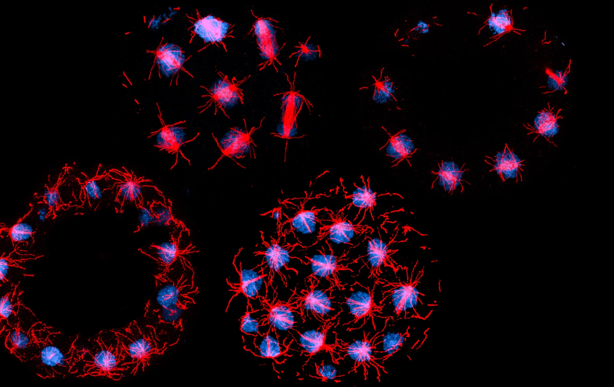
The first species that Dudin’s lab imaged using expansion microscopy was the unicellular eukaryote Sphaeroforma arctica, a protist that has multiple nuclei in a single cell. This image shows cells at different stages of their life cycle. Nuclei in the two upper cells are undergoing mitosis, or nuclear division. The lower cells are in late mitosis, with the nuclei switching back to a growth stage.
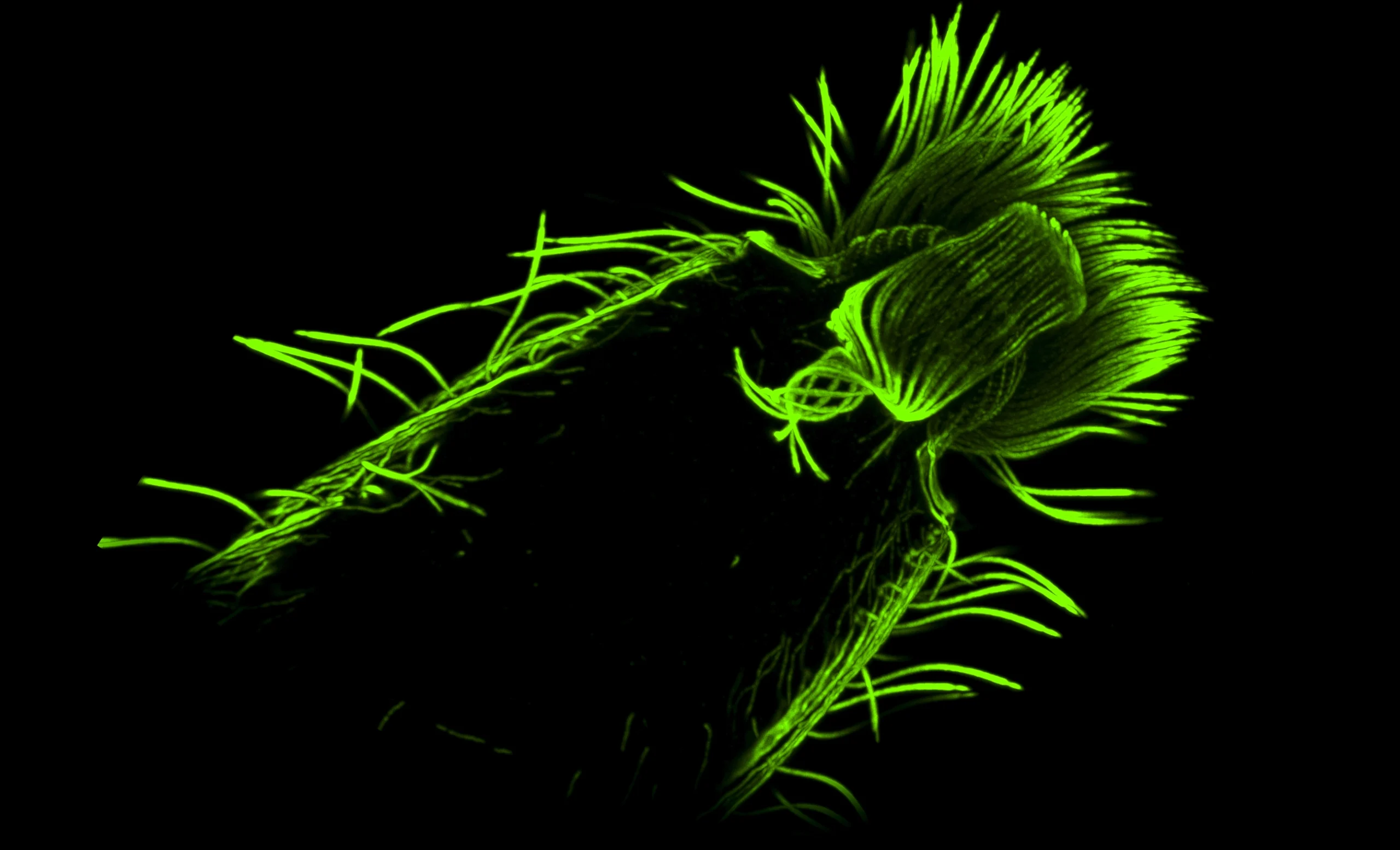
The predatory ciliate Lacrymaria is a Dudin lab favorite and has a feeding structure formed from a vortex of microtubules. Although it is retracted here, Lacrymaria can rapidly extend the tubule structure to capture escaping prey.
Expansion microscopy has allowed Dudin and Dey to see all sorts of unexpected structures. For instance, Rhinomonas is a unicellular photosynthetic alga with two flagella, found predominantly in marine environments. The appearance of the specialized protein centrin (green) decorating regularly spaced arrays was previously unreported for this lineage.
Diatoms are particularly difficult to image through their protective glass shells, called frustules. Dey’s lab decided to use an alternative expansion microscopy protocol, in which the sample is expanded and its proteins tagged with fluorescent antibodies; rapid freezing then locks everything in place.
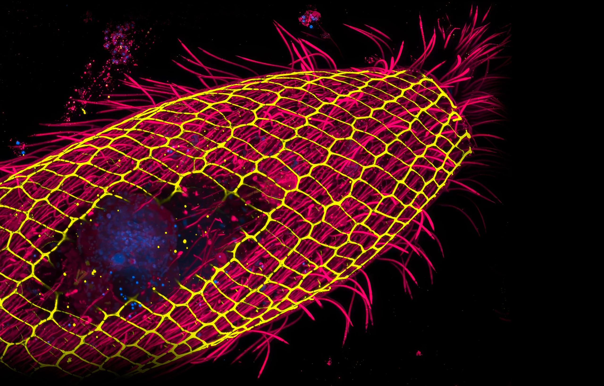
Centrin is a small calcium-binding protein that co-polymerizes with larger proteins to produce networks of structural filaments in a cell. Centrin-containing fibers are especially abundant in ciliates; this intricate mesh (yellow) is located just below the cell membrane in this expanded Vorticella cell.
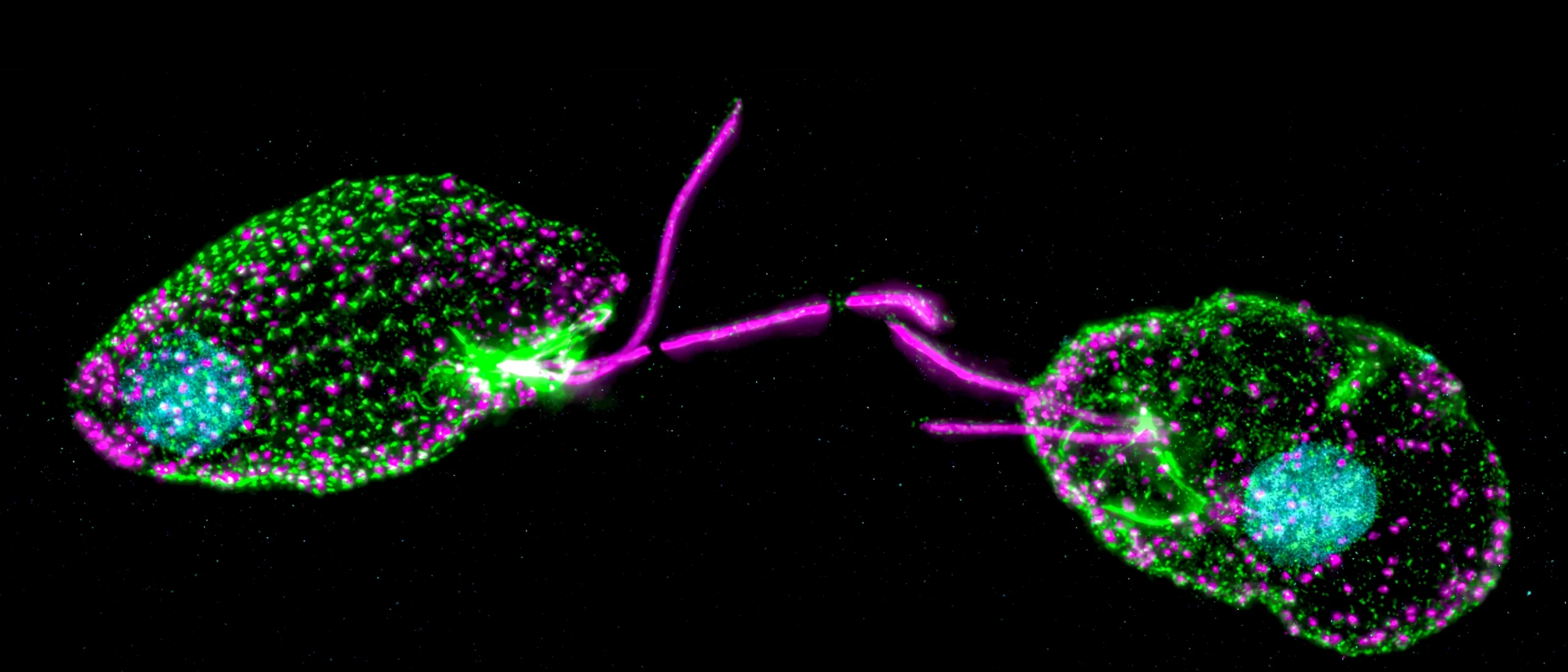
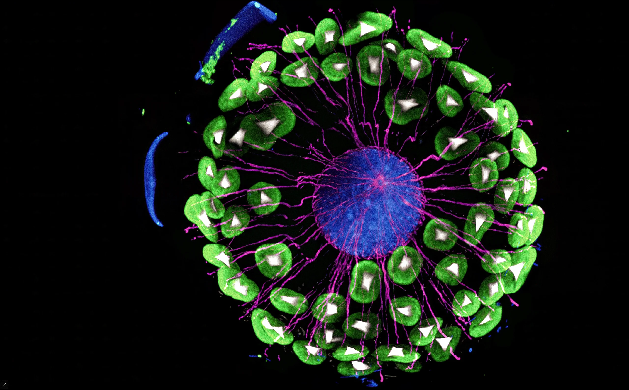
In freshwater ciliates known as Vorticella, the researchers spotted “an amazing centrin cage, dotted tubulin flagellar components, and an insane mouth apparatus [at far left, green],” Dudin said. In the middle, a spindle (green) appears to be organizing as cells prepare for division. Green dots of the structural protein tubulin are visible throughout the protist’s distinctive stalk (at right).
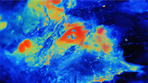
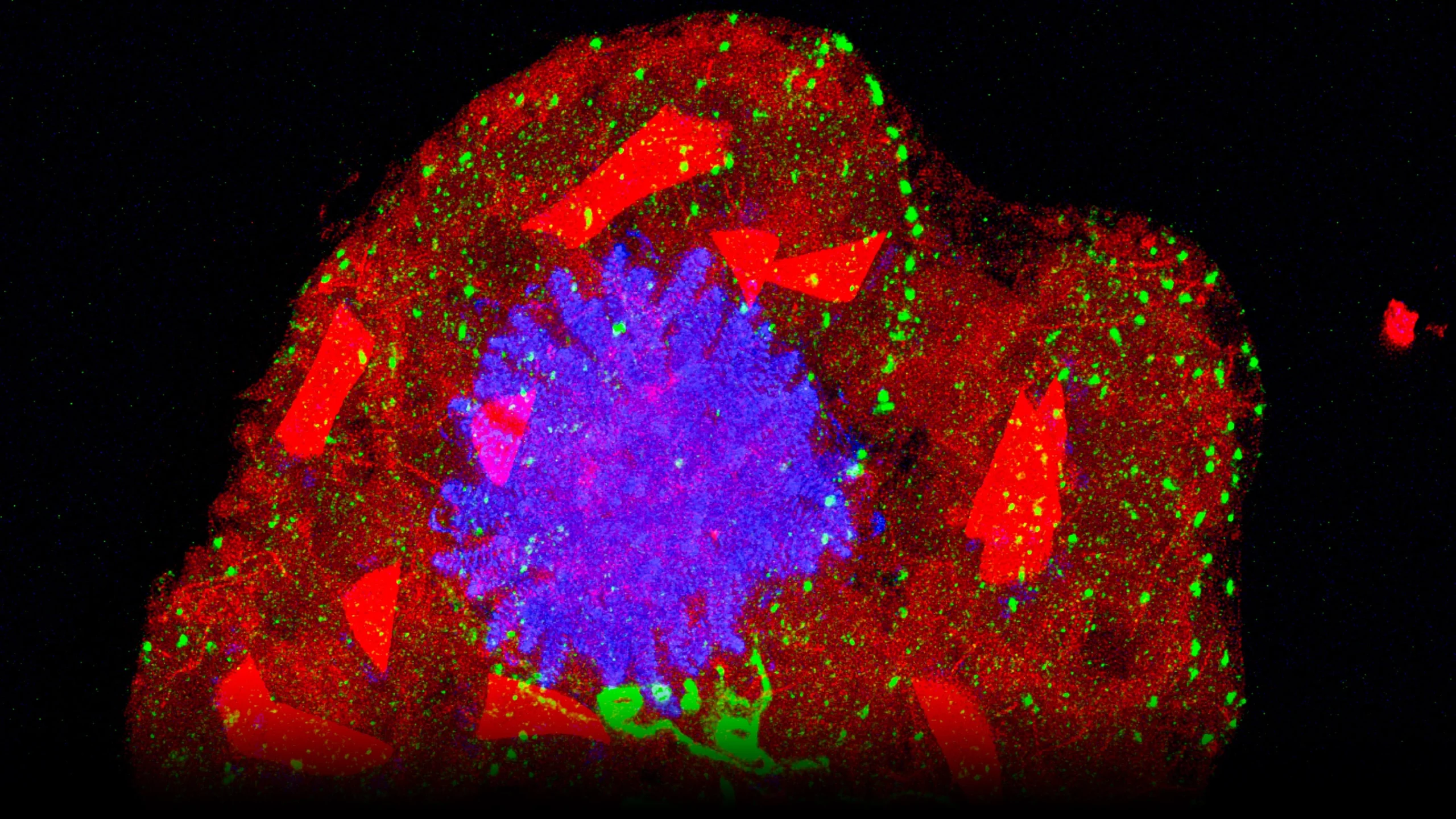
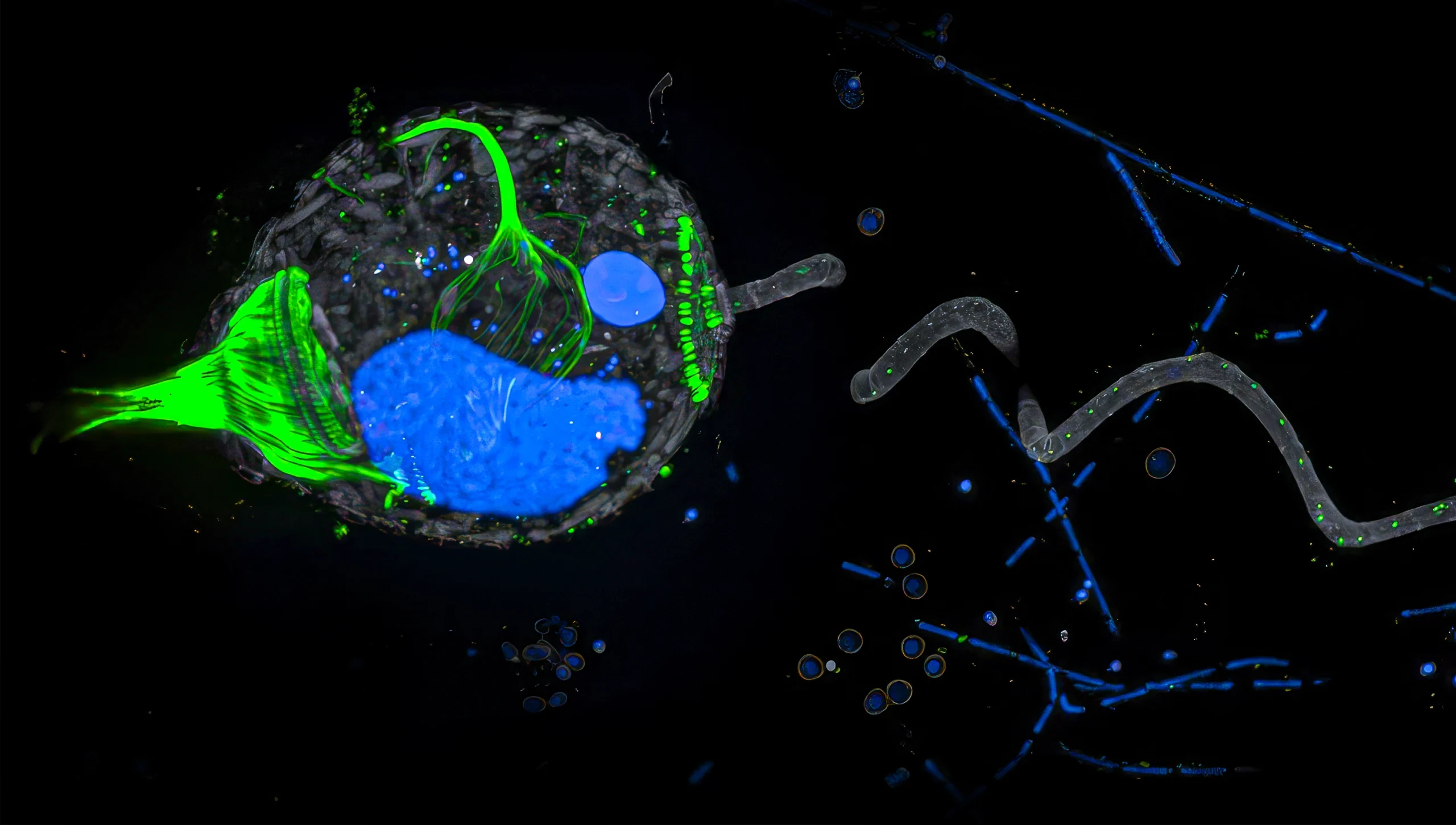
A dinoflagellate identified in Antarctica’s Ross Sea is known to steal organelles called plastids from the microalga Phaeocystis antarctica. Expansion microscopy of the engulfing process revealed that the host microbe retained more than just plastids. The alga’s nucleus (blue) and other features such as a protein complex crucial for photosynthesis (green) were also observed inside the host. These interactions offer insight into early evolutionary steps of eukaryotes but are still not fully understood.
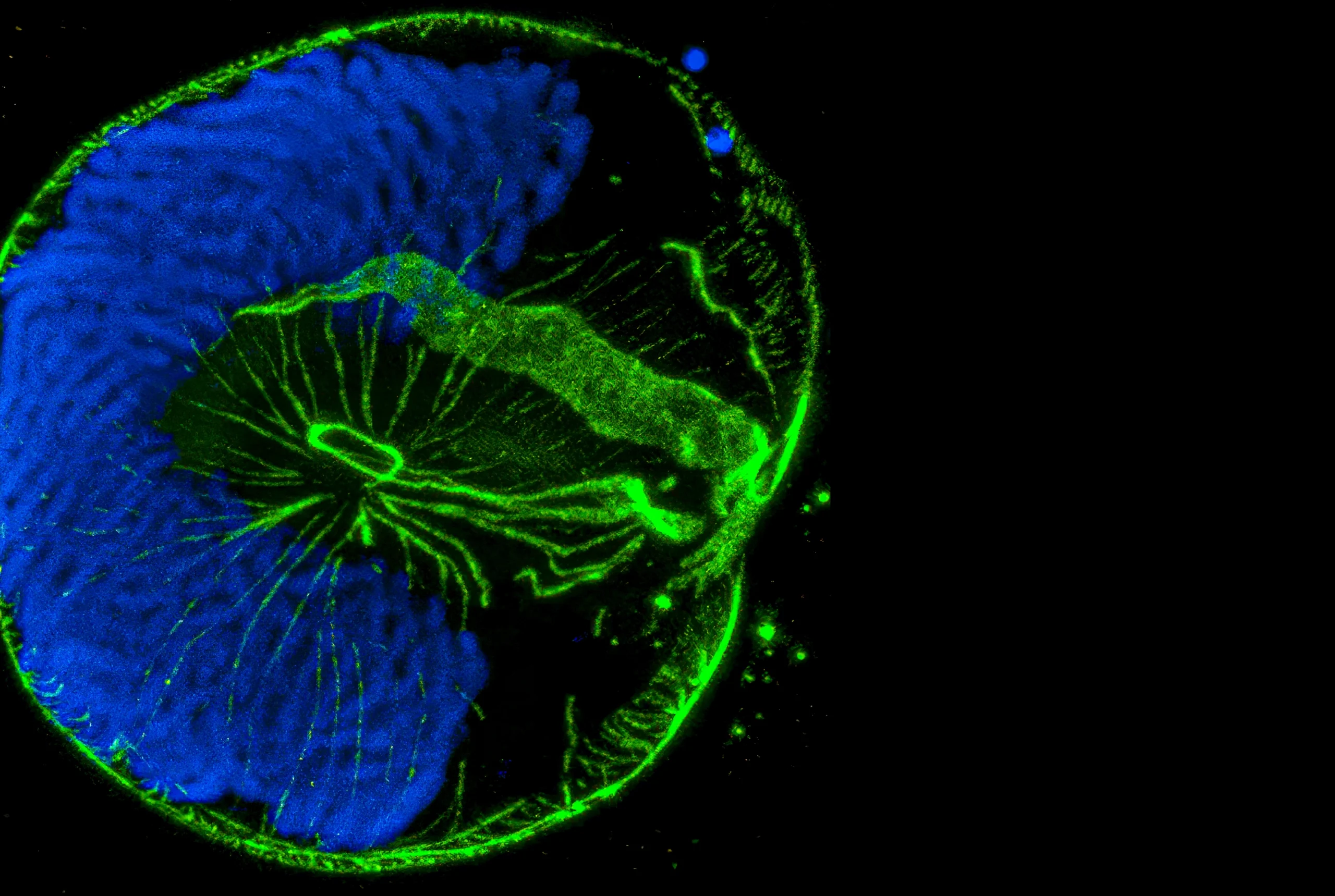
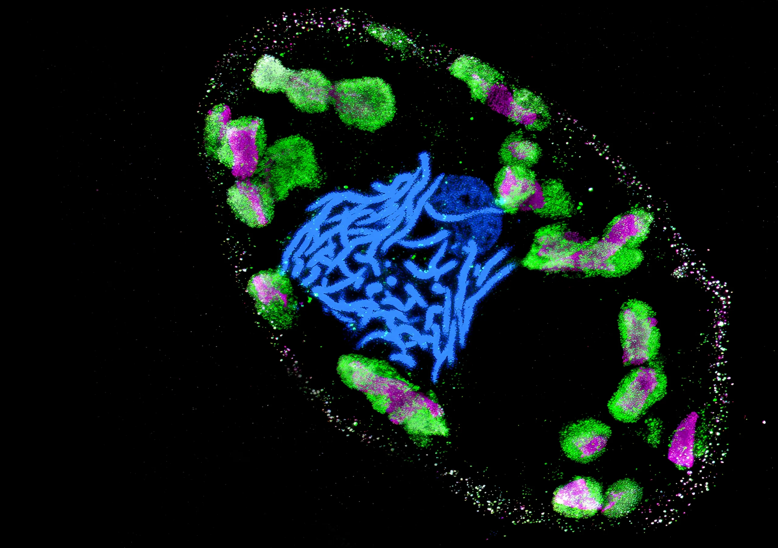
Expansion microscopy of the dinoflagellate Lingulodinium polyedra reveals the chromosomal organization (DNA in blue) and internal axostyle (center, green) — a microtubule structure that helps the cell move. “We have imaged more than 60 species across the dinoflagellate tree, allowing us to map cytoskeletal diversity within the whole lineage,” Dudin said.