Cells Use ‘Bioelectricity’ To Coordinate and Make Group Decisions
Introduction
We’re used to thinking of the brain as an electric organ. The rest of the body? Not so much. But it would be a mistake to dismiss your other tissues as dumb hunks of electrically inert flesh. Even the protective layers of cells that compose your skin and line your organs use electrical signals to make decisions, according to recent research.
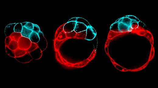
Results published in Nature show that cells use bioelectricity to coordinate a complex collective behavior called extrusion, a vital process that ejects sick or struggling individual cells from tissue to maintain health and keep growth in check. Merciless as it might seem, extrusion helps keep you alive. It’s vital for the health of protective epithelial tissues, and when it goes wrong, it can contribute to disease, including cancer and asthma. Until now, it’s been unclear how cells were singled out for this process.
According to the new results, as epithelial tissue grows, cells are packed more tightly together, which increases the electrical current flowing through each cell’s membrane. A weak, old, or energy-starved cell will struggle to compensate, triggering a response that sends water rushing out of the cell, shriveling it up and marking it for death. In this way, electricity acts like a health checkup for the tissue and guides the pruning process.
“This is a very interesting discovery — finding that bioelectricity is the earliest event during this cell-extrusion process,” said the geneticist GuangJun Zhang of Purdue University, who studies bioelectrical signals in zebra fish development and wasn’t involved in the study. “It’s a good example of how a widening electronic-signaling perspective can be used in fundamental biology.”
The new discovery adds to the growing assortment of bioelectrical phenomena that scientists have discovered playing out beyond the nervous system, from bacteria swapping signals within a biofilm to cells following electric fields during embryonic development. Electricity increasingly appears to be one of biology’s go-to tools for coordinating and exchanging information between all kinds of cells.
“People just kind of relegated [bioelectricity] to ‘This is just neurons.’ No — it’s all of our bodies,” said study author Jody Rosenblatt, an epithelial cell biologist at King’s College London and the Francis Crick Institute. “There are electrical currents going through your body all the time, and they’re doing things.”
Life’s Spark
It’s no coincidence that Frankenstein’s monster sprang to life with a spark. In the late 18th century, just a few decades before Mary Shelley wrote her science fiction masterpiece, the Italian surgeon Luigi Galvani jolted the scientific community with experiments that used metal and electricity to compel disembodied frog legs to kick. He became convinced that there was an “animal electricity” running through life.
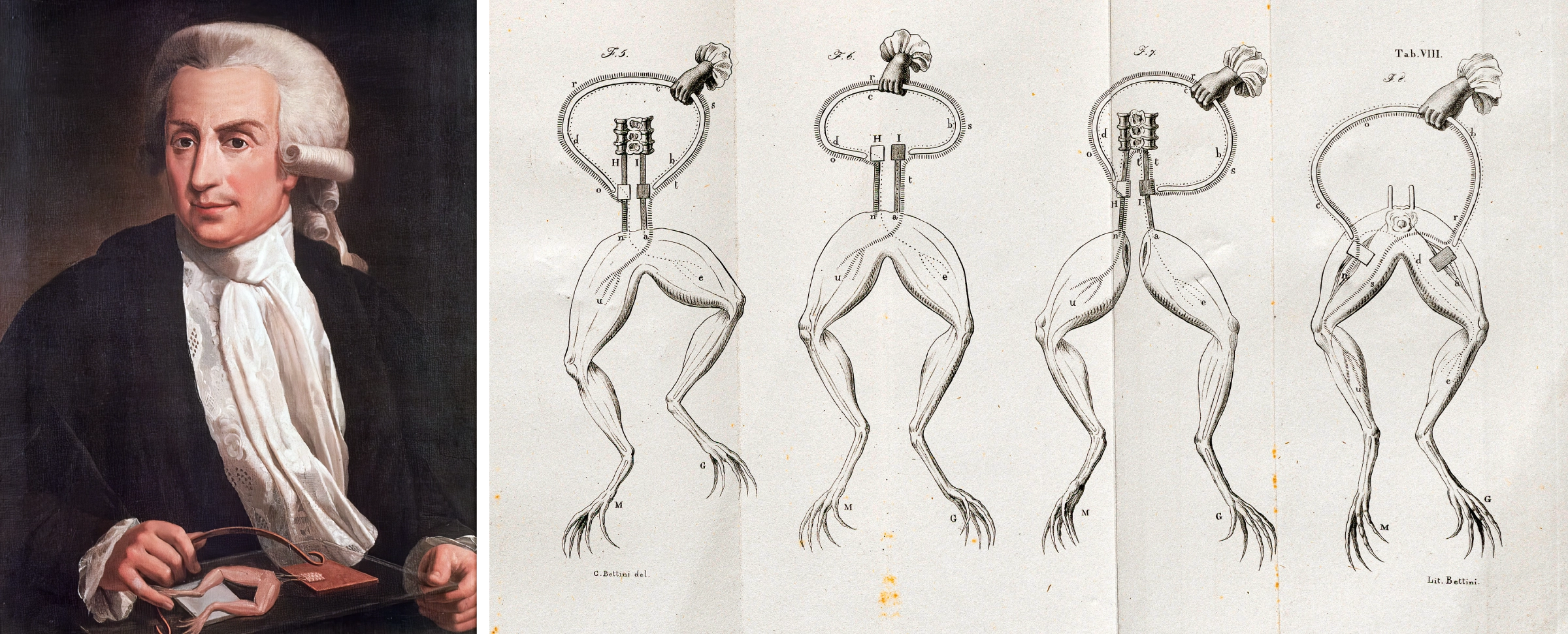
In the late 1700s, the anatomist Luigi Galvani (left) discovered that dissected frog legs twitched when touched with an electrical spark, setting off the study of what he called “animal electricity.”
Public Domain; ETH-Bibliothek Zurich / Science Source
While Galvani was later proven wrong in the details, he wasn’t totally off. Virtually every cell on every branch of the tree of life expends a hefty chunk of its energy budget — in some cells, more than half — on maintaining a voltage across its membrane. The voltage difference that results, called the membrane potential, stores potential energy that can be released later. It’s like the pressure behind a dam: Gravity tugs water downhill, and dams store energy by holding water at the top of a hill. Like gravity, the electrochemical force tugs charges “downhill” — positive charges stream toward negative charges and vice versa in electrical currents. Blocking that flow, for example with a cell membrane, stores up electrical potential energy.
The electric currents that pour from our wall sockets are streams of electrons. In cells, “it’s quite similar, but not exactly the same,” said Elias Barriga, who studies tissue biophysics at the Dresden University of Technology. “We are fueled by ions.”
Ions are atoms or molecules that carry charge because of extra or missing electrons, which give them negative or positive charges, respectively. They can enter and exit cells only through specialized protein channels and pumps. Just as hydroelectric plants can use surplus energy to pump water back up into the reservoir for later use, cells use their chemical energy to pump ions “uphill” against the electric flow. By controlling the natural current and letting positive or negative charge build up on either side of their membranes, cells maintain their membrane potential. And if this energy is used or leaks away, cells can replenish it by expending more of their chemical energy.
“You generate a potential: what’s inside versus what’s outside, a different concentration of ions,” Barriga said. “That is the source of bioelectricity.”
Neurons make use of this biological electricity to share information. By releasing messenger molecules called neurotransmitters that open and close ion channels, neurons can nudge their neighbors’ membrane potentials up or down. If these chemical nudges push a neuron’s membrane potential beyond a threshold, the cell “spikes” — voltage-sensitive ion channels throw open the floodgates for positive sodium ions, which rush into the cell and cause a rapid voltage swing that ripples along the neuron’s length. When that signal reaches the interface between neurons, voltage-sensitive channels open wide, triggering the release of neurotransmitters to more neurons downstream.
Muscle contraction also kicks off with a voltage spike; neurons send electrical signals streaming into muscle fibers, triggering contractions. This is why Galvani’s electrified frog legs twitched, and why a jolt of electricity can jump-start a stopped heart. (Specialized cells in the heart use electricity to set the pace of its regular contractions.) While all tissues maintain membrane potentials, researchers don’t really know what they do. Compared to electrophysiology, which often focuses on electricity in the brain and heart, the field of bioelectricity — a grab-bag term for electrical activity everywhere else in organisms — has lagged behind, Barriga said.
“I think that at some point it got stuck,” he said. “But now I can tell you that that is coming back like crazy.”
A Shocking Discovery
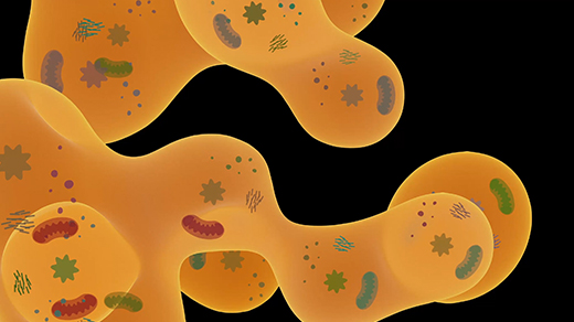
The epithelial tissues that make up skin and line organs, blood vessels, and body cavities quietly burn about 25% of their available energy to maintain membrane voltages between minus 30 and minus 50 millivolts. But researchers interested in these tissues typically study mechanical forces, chemical signaling, and gene expression — not currents and voltage, Rosenblatt said.
Until recently, that included her. Rosenblatt has spent 25 years piecing together the details of epithelial extrusion, a process that keeps tissue growth in check. Because epithelial cells grow quickly, even a slight mismatch between the rates of cell division and cell death can quickly add up to a tumor or injury. Runaway replication can grow into cancer, while overzealous culling — as can happen in asthma — compromises the integrity of tissues. It’s important to get the balance right.
Around 14 years ago, Rosenblatt and colleagues discovered that overcrowded epithelial cells are popped up and out of the tissue alive — extruded — to maintain that tissue balance as new cells grow. That raised a question: How does tissue “choose” which living cells to expel?
In earlier work, Rosenblatt’s team watched as some cells dumped their water and shriveled up like raisins before being extruded; indeed, this shrinkage seemed to kick off the process. But the researchers didn’t know what caused the cells to shrink in the first place. They didn’t work on bioelectricity and were unaware of any effect it might have.

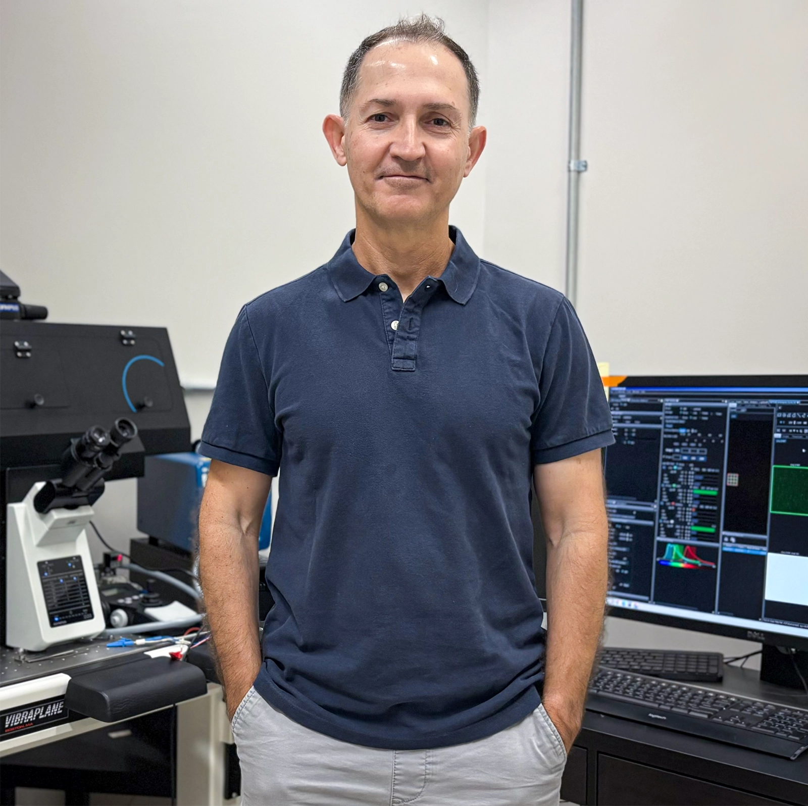
The cell biologist Jody Rosenblatt studies extrusion, the process by which a tissue expels cells to prevent overcrowding. Her lab recently described bioelectricity as helping the tissue “choose” which cells to extrude.
Antonio Tabernero
In further experiments, they were able to prevent the cells from shrinking by blocking a pressure-sensitive ion channel in the cell membrane that opens when squeezed. They decided to see if blocking other ion channels might interfere with extrusion too.
“We got so many hits, we were just like: Jesus, this is crazy,” Rosenblatt recalled. One of those hits was a voltage-gated potassium channel, like those that open up during a neuron’s voltage spike. It struck Rosenblatt as “weird” enough to follow up on. Using special dyes that reveal the voltage across cell membranes, the scientists found that epithelial cells destined for extrusion — and only those cells — lose their membrane potential about five minutes before shrinking and being extruded. The result was clear: Extrusion kicks off with an electrical signal.
Instead of sending neurotransmitters back and forth like neurons, densely packed epithelial cells squeeze each other. As the tissue gets more crowded, the squeezing intensifies. This opens pressure-sensitive ion channels, which allow positive sodium ions to leak across the squashed cells’ membranes and into the cell.
Faced with this challenge, a healthy cell will use its chemical energy to activate pumps to push sodium back out and restore its normal voltage. But stressed or unhealthy cells without energy to spare can’t keep up. Their membrane voltage falters, throwing open those “weird” voltage-sensitive channels. When that happens, water pours out of the cell in a “lightning” flash clearly visible in microscope images, Rosenblatt said. Once a cell loses 17% or more of its volume, it is extruded. Her working hypothesis is that a biochemical cascade set off by the shrinkage contracts motor proteins, which mechanically extrude the cell.
In this way, bioelectrical flow across cell membranes lets tissues test which cells are the least healthy and mark them for extrusion. “They’re always pushing against each other and bullying each other. And what they’re doing is probing each other for which one’s the weakest link,” Rosenblatt said. “It’s a community effect.”
Evolution as Electrician
At the University of California, San Diego, the biophysicist Gürol Süel studies electricity in bacterial biofilms, which are collectives composed of single-celled bacteria that can also survive independently. The signaling that Rosenblatt and her team described in human tissues has several things in common with electrical mechanisms Süel has described in microbes — and which appear again and again across the tree of life.
“It’s a very elegant study, very nice results,” he said of the new research. “And conceptually, it makes sense.”
Gürol Süel studies how bacteria in biofilms use bioelectricity to communicate and coordinate. A membrane potential “tells you, in one glance almost, about the state of the cell,” he said.
Suel Lab
Electricity increasingly appears to be one of evolution’s go-to solutions for integrating multiple streams of information. Epithelial tissues use it to keep tabs on crowding. Neurons compile input signals from multiple sources into a spiking output. A Venus flytrap snaps shut when sensory hairs with touch-sensitive ion channels react to prey. These channels are tuned to trigger a voltage spike and tell the trap to close only if stimulated multiple times in rapid succession.
“The membrane potential is so fundamental, and it is very fast,” Süel said. While switching genes on and off or upping protein production could take minutes or hours, a membrane potential can flip in fractions of a second. “It tells you, in one glance almost, about the state of the cell,” he added.
Ten years ago, Süel and his team showed that microbes in biofilms can spike their membrane potentials to communicate, just as neurons do. Since then, they’ve shown that biofilms use electricity to coordinate tasks, prevent runaway growth, and invite free-swimming bacteria to join the collective. Bioelectricity can even help them avoid falling victim to the tragedy of the commons: Two biofilms sharing scarce food can send electrical signals to each other to take turns eating.
Multicellular animals, too, use electrical signaling to organize themselves. Zhang, of Purdue, studies bioelectrical signaling in zebra fish, which develop striking extra-long tails when a certain ion channel is mutated. This suggests that electrical signaling somehow tells tissues in a developing embryo how long to grow. Michael Levin, a researcher at Tufts University, has blocked cell channels to manipulate the membrane potentials of developing worm embryos, causing genetically identical worms to develop different body plans. And last year, Barriga and his colleagues showed that frog embryos generate natural electric fields that guide the migration of specific stem cells to their proper locations in the developing embryo.
The failure of bioelectric processes might be an overlooked cause of disease. Cancerous cells tend to have different membrane potentials than healthy ones, and Levin has argued that some cancers might result from a breakdown in multicellularity that happens when cells can no longer use electricity to coordinate. For example, maybe they can no longer communicate the message “I’m struggling and should be extruded,” and the result is the uncontrolled growth and, ultimately, a tumor.
Süel is convinced that bioelectricity is as old as life itself. Indeed, an electric current drives the molecular turbines that synthesize life’s universal energy currency, ATP, in every cell alive today. One leading origin-of-life scenario places the beginning at deep-sea hydrothermal vents. There, natural currents of positively charged protons could have served as a kind of primordial membrane potential and powered prebiotic chemical reactions. But whether life started with such a spark or not, bioelectricity’s ubiquity suggests it has deep evolutionary roots that we’re just beginning to unearth.
“There are a lot of interesting things that cells are probably doing, just like this paper showed, that we just don’t know yet,” Süel said. “We have not uncovered even half of this. … There’s a lot of opportunity for discovery.”