Once Thought To Support Neurons, Astrocytes Turn Out To Be in Charge
Introduction
The human brain is a vast network of billions of neurons. By exchanging signals to depress or excite each other, they generate patterns that ripple across the brain up to 1,000 times per second. For more than a century, that dizzyingly complex neuronal code was thought to be the sole arbiter of perception, thought, emotion, and behavior, as well as related health conditions. If you wanted to understand the brain, you turned to the study of neurons: neuroscience.
But a recent body of work from several labs, published as a trio of papers in Science in 2025, provides the strongest evidence yet that a narrow focus on neurons is woefully insufficient for understanding how the brain works. The experiments, in mice, zebra fish, and fruit flies, reveal that the large brain cells called astrocytes serve as supervisors. Once viewed as mere support cells for neurons, astrocytes are now thought to help tune brain circuits and thereby control overall brain state or mood — say, our level of alertness, anxiousness, or apathy.
Astrocytes, which outnumber neurons in many brain regions, have complex and varied shapes, and sometimes tendrils, that can envelop hundreds of thousands or millions of synapses, the junctions where neurons exchange molecular signals. This anatomical arrangement perfectly positions astrocytes to affect information flow, though whether or how they alter activity at synapses has long been controversial, in part because the mechanisms of potential interactions weren’t fully understood. In revealing how astrocytes temper synaptic conversations, the new studies make astrocytes’ influence impossible to ignore.
“We live in the age of connectomics, where everyone loves to say [that] if you understand the connections [between neurons], we can understand how the brain works. That’s not true,” said Marc Freeman, the director of the Vollum Institute, an independent neuroscience research center at Oregon Health and Science University, who led one of the new studies. “You can get dramatic changes in firing patterns of neurons with zero changes in [neuronal] connectivity.”
Astrocytes do not engage in the rapid-fire signaling typical of neurons at synapses. Instead, they monitor and tune higher-level network activity, dialing it up or down to maintain or switch the brain’s overall state. This function, termed neuromodulation, may cause an animal’s brain to switch between dramatically different states, such as by gauging when an action is futile and prompting the animal to give up, one of the new papers shows.
Neuromodulation is necessary for keeping the brain’s activity level in a functional range, preventing it from either flatlining or erupting in seizures. “No neural circuit would work at all without continual fine-tuning by these things we call neuromodulators, [the molecules that mediate the adjustments],” said Stephen Smith, an emeritus professor of neuroscience at Stanford University who conducted pioneering experiments in astrocyte signaling in the late 1980s and early 1990s and was not involved in the new research.
For many years, that fine-tuning was thought to be conducted by neurons themselves. While previous work has implicated astrocytes in some cellular signaling, the latest experiments use “advanced techniques to really pinpoint and satisfy beyond a doubt that astrocytes are having a key role in neuromodulation in the brain,” said Douglas Fields, an emeritus neuroscientist at the National Institutes of Health who was not involved in the new research.
In that role, astrocytes could be major participants in sleep or psychiatric disorders that broadly disrupt the state of the brain. “We have to think about what this means for neuropsychiatric disease,” Freeman said.
A Star Is Born
Astrocytes are a type of glial cell, a class of non-neuronal nervous system cells that tile the brain, filling the space between neurons like packing peanuts. Greek for “glue,” the name “glia” reflects the mid-18th-century idea that the cells’ purpose was simply to hold the brain together.
By the 1950s, researchers knew that astrocytes did more than that. In experiments, the cells sucked up excess neurotransmitters, buffered potassium, and secreted substances that neurons require for energy. Like cellular alchemists, astrocytes seemed to be monitoring and adjusting the broth of the brain, keeping conditions favorable for neurons. But scientists considered them relatively passive regulators until the late 1980s, when Smith built a new microscope for his neuroscience lab at Yale University.

The neuroscientist Stephen Smith, pictured here with Banjo the Havanese dog, built a microscope in the late 1980s that prompted pioneering research into astrocyte signaling.
Lyn Flaim Healy
Smith’s novel digital video fluorescence microscope was designed to take movies of neuronal activity using fluorescent light. When a neuron fires, calcium rushes into the cell. So the researchers put fluorescent sensors into brain cells that glowed when they encountered calcium. The microscope could detect the light as it brightened and dimmed over space and time, revealing the cells’ firing patterns. “We had probably the most advanced, sensitive, coolest setup going,” Smith said.
One day in 1989, Smith’s graduate student Steve Finkbeiner (now a neurologist at the nonprofit Gladstone Institutes in San Francisco) was using the microscope to explore the potentially toxic effects of the neurotransmitter glutamate, the molecule most neurons in the brain use to communicate. Finkbeiner was not interested in astrocytes, but because they help keep neurons alive, he put them in his cell culture. Then he added glutamate.
“He’s all of a sudden yelling and screaming from his microscope setup: ‘Hey boss, come here! You’ve got to see this!’” Smith recalled. “They [the astrocytes] went completely nuts.” Fluorescence rippled across the bed of astrocytes in waves, hopping from one cell to the next. These calcium waves showed coordinated activity, as if the astrocytes were communicating with each other. And because the cells responded to glutamate, it was only logical that they would also respond to neurons. In their 1990 paper describing the experiment, the researchers boldly proposed that “networks of astrocytes may constitute a long-range signaling system within the brain.” Other teams soon showed that astrocytes in dishes, brain slices, and even anesthetized animals responded to various neurotransmitters.
Many neuroscientists at the time likened astrocytes’ newfound properties to those of neurons, but in retrospect the differences seem glaring. For one thing, astrocytes occupy relatively massive territory: One astrocyte covers a large expanse of tissue, reaching as many as 2 million synapses in the human brain. Astrocytes work on longer timescales than neurons do. Their calcium waves spread over a period ranging from seconds to minutes — much longer than the milliseconds it takes for neurons to propagate signals down their axons and release neurotransmitters.
To study how this surprising new view of astrocytes related to behavior, research groups turned to animal models. Researchers tried to activate astrocytes in lab mice by bombarding them with sensory stimuli, such as by shining light in their eyes or touching their whiskers; they looked for a response through a cranial window under a fluorescent microscope. Sometimes the cells responded, sometimes they didn’t. Then, in 2013 and 2014, two independent research teams reported a sure-fire way to get astrocytes’ attention: They startled the mice by surprising them with a puff of air or by abruptly turning on a treadmill under their feet. The startle response is a largely unconscious defense mechanism and a sudden switch in brain state, found throughout the animal kingdom.
When vertebrate animals are startled, neurons in a brainstem region called the locus coeruleus release norepinephrine, a neuromodulator associated with arousal, along fibers that fan out across the brain. Instead of sending a specific message, as neurotransmitters do, neuromodulators dial brain activity up or down and change the brain’s overall state like a dial on a radio. The studies indicated that norepinephrine was the trigger for the astrocyte waves, implicating astrocytes in neuromodulation in some capacity.
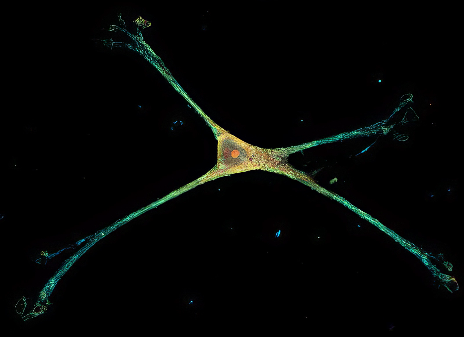
A single rat astrocyte is splayed on a specialized nanowire structure. In its native environment, the cell would envelop hundreds of thousands of synapses, enabling it to monitor and adjust neuronal signaling.
Johns Hopkins University
Still, so much about astrocyte signaling remained mysterious. The cells were known to have norepinephrine receptors, but no one knew how the binding of norepinephrine led to the calcium waves. And there was still the question of what signal those waves sent to downstream neurons. Some researchers thought astrocytes produced their own “gliotransmitter” molecules that acted upon neurons, but others disputed that notion. At meetings, researchers engaged in loud, heated debates over how much — indeed, whether — astrocytes shape the flow of information in the brain.
A student in Freeman’s lab, Zhiguo Ma, then at the University of Massachusetts Medical School, sought to settle the issue in a fruit fly brain. “Please don’t,” Freeman recalled warning him. “It’s such a mess.” Ma forged ahead. He replicated the startle response in fruit flies by suddenly flipping them upside down. Using the delicate tools of molecular biology, he traced the chemical relay: The fly versions of norepinephrine activated astrocytes by opening a channel in the cell membrane, causing the release of a gliotransmitter — likely adenosine — that squelched neuronal signaling. It was critical to characterize such neuron–astrocyte interactions, “as they would represent a potentially widespread mechanism for controlling brain function,” Freeman’s team wrote in Nature in 2016.
To some, the experiment provided the first proof that astrocytes are integral parts of neural circuits. But one fruit fly paper was not enough to sway skeptics. Nearly a decade later, eerily parallel findings in a vertebrate would tip the scales.
When To Give Up
Although we don’t often think of it this way, the act of giving up reflects a sudden shift in brain activity. It represents a change in mental state from hope to hopelessness that, like being startled, has profound effects on behavior. Researchers led by the neuroscientist Misha Ahrens were studying what made zebra fish larvae give up when they made a discovery about how astrocytes mediate such a sudden change in mood.

Misha Ahrens showed that astrocytes modulate the switch in brain state from hope to hopelessness — specifically, to the state of giving up after engaging in a futile effort.
© HHMI, photo by Toby Hayman
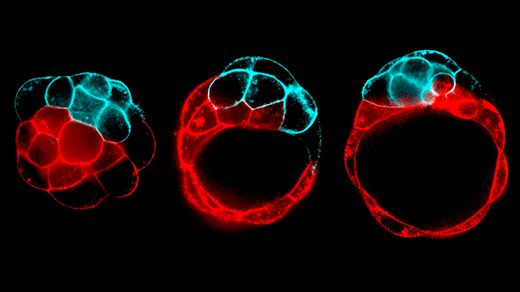
What does it look like when a zebra fish gives up? In the wild, if a zebra fish wants to stay put in flowing water, it will swim against the current. In the lab at the Howard Hughes Medical Institute’s Janelia Research Campus in Virginia, Ahrens’ team used virtual reality to create a simulation of a current in the zebra fish tank, so that a fish would think it was slipping backward no matter how furiously it swam. The fish would swim harder at first, but after about 20 seconds, it would typically give up. A little while later, it would try again.
All the while, the researchers monitored neurons and astrocytes in the zebra fish’s brain using advanced whole-brain imaging techniques. As the fish fruitlessly fought the current, neurons that release norepinephrine fired; in response, calcium built up in astrocytes. The buildup paralleled the number of attempts the fish made to fight the current, as if the astrocytes were keeping track — until at some point they issued a stop signal, and the zebra fish gave up.
When Ahrens’ team disabled the astrocytes using a laser, the fish never stopped swimming. And if the astrocytes were artificially activated, the fish stopped right away. “It was the first time that it was shown that astrocytes had a role in behavioral state switching,” Ahrens said.
In an ensuing Science paper, published in 2025, the researchers revealed how astrocytes caused these changes in behavior. Using fluorescent sensors for various molecules, they found that when enough calcium builds up in astrocytes, they release the energy molecule ATP, short for adenosine triphosphate. Outside the cell, the ATP is converted into adenosine, which acts on neurons — in this case, by exciting neurons that inhibit swimming and suppressing swim neurons. This sequence echoes what Ma and Freeman observed in the fruit fly.
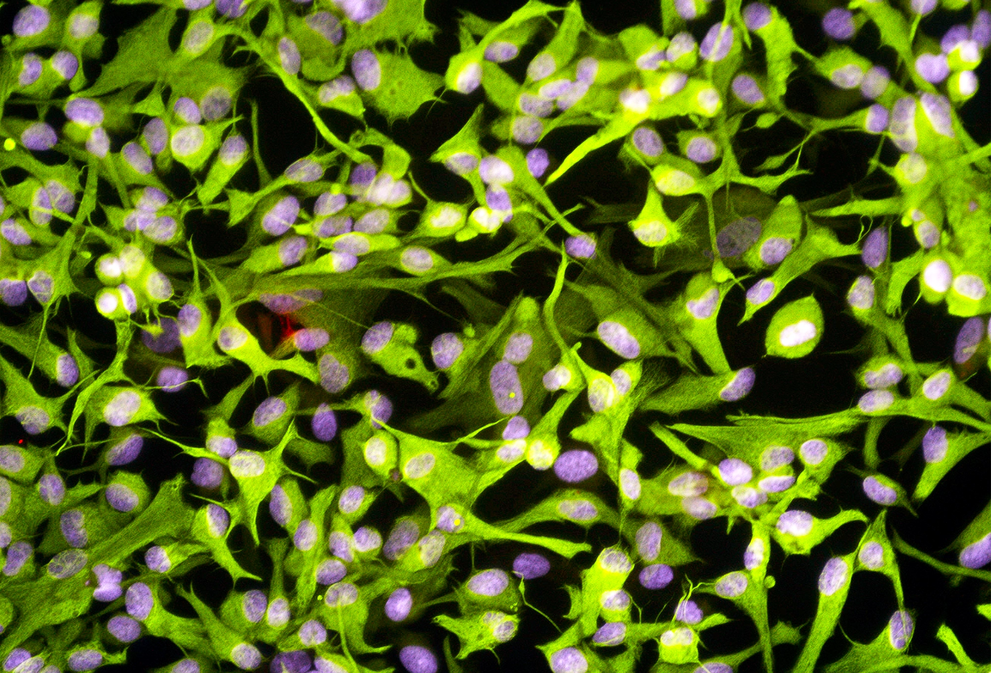
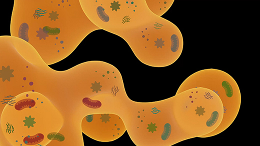
For a long time, astrocytes (seen here in a fluorescence light micrograph of human tissue) were considered mere support and scaffolding for all-important neurons. The new experiments reveal in great detail the cells’ influence over neuronal signaling in the brain.
Science Source
The same molecular chain of events also showed up in the mouse brain, according to research led by Thomas Papouin at Washington University School of Medicine and published in the same Science issue. Papouin’s team was studying changes at synapses that alter communication between neurons, a form of neuroplasticity that underlies ongoing shifts in thought and behavior. Norepinephrine was thought to produce these shifts by acting directly on neurons. But to Papouin’s surprise, norepinephrine’s effects were apparent even when its receptors on neurons had been removed. The process depended solely on astrocytes.
“We did expect that, in large part, the effect of norepinephrine on synapses would be mediated by astrocytes,” Papouin said. “But we did not expect all of it to be!”
The finding of parallel molecular pathways in such distinct species as fruit flies, zebra fish, and mice points to “an evolutionarily conserved way in which astrocytes can profoundly affect neural circuits,” Freeman said.
The results suggest a gaping hole in previous theories of neuromodulation. “In the past, neuroscientists studied neuromodulators and knew they were important in regulating neural circuit function, but none of their thinking, none of their diagrams, none of their models had anything in them other than neurons,” Fields said. “Now we see that they missed a big part of the story.”
Thomas Papouin discovered that a type of neuroplasticity, which underlies shifts in thought and behavior, is mediated entirely by astrocytes, neurons not required.
Courtesy of Thomas Papouin
The fruit fly research by Freeman’s team indicates the next steps in research in vertebrates; in the same Science issue the group reported that norepinephrine changes how astrocytes respond to input from neurons. Freeman’s postdoc Kevin Guttenplan doused a dissected fly brain with the fly versions of norepinephrine. “All of a sudden, the astrocytes went from responding to none of the other neurotransmitters to responding to all of them,” Guttenplan said. Norepinephrine and its analogues in the fly seem to enable astrocytes to “hear” neurons’ molecular messages and then modulate their activity.
This dynamic helps explain how astrocytes can rapidly switch the brain from one state to another. “If there’s low norepinephrine, which would mean low arousal, astrocytes don’t listen much at all to other synapses,” Freeman said. “But as soon as you arouse the animal, and there is norepinephrine around, now astrocytes can listen to every synapse, and they can turn around and change how neurons fire in response to that.”
The results reveal a new complexity to how the brain processes information, Guttenplan said. “On top of the already complicated connectome [network of neurons], you have this whole other layer of regulation.”
Mood Meter
Although the details of astrocytes’ signaling mechanism are coming into focus, they still lag behind what is known about neurotransmission. “It’s an exciting time,” said Alex Chen, a student at Harvard Medical School and first author of the zebra fish paper. “For the astrocyte field, at least conceptually, we are not very far ahead of where people were for neurons” at the onset of modern neuroscience in the 1950s.

Working in zebra fish, Alex Chen observed the first known instance of astrocytes mediating a rapid transition between brain states.
Courtesy of Harvard_MCB
Meanwhile, researchers are homing in on critical brain functions that astrocytes mediate. Some research suggests that astrocytes’ ability to accumulate information over time (as happened with the zebra fish’s swim attempts) extends to the sleep-wake cycle. Astrocytes appear to keep track of people’s increasing sleep debt throughout the day, likely through a buildup of calcium, and secrete sleep-inducing molecules that alter brain activity.
“We see astrocytes implicated in behaviors associated with big state transitions — like sleep, hunger, arousal — where you need multiple types of circuits over a very large area to get turned on and off, especially on a slower timescale,” Guttenplan said.
Those behaviors may reflect mental health conditions. Last year, researchers revealed a neuron-astrocyte brain circuit that was triggered by stress and produced behavior resembling depression in mice. It’s possible that some mental health disorders are disorders of astrocyte signaling. People’s moods change relatively slowly, Ahrens said, in a process partly driven by neuromodulators. Astrocytes’ role in neuromodulation points to their promise as a drug target.
“Neuroscience has only cared about neurons for a century now, and we don’t yet have a cure for a single brain disorder,” Papouin said. The way to change that, he said, is to accept the existence and influence of non-neuronal cells such as astrocytes, and to include them in models and experiments.
Most neuroscientists haven’t received that memo, Freeman said. “Ninety-nine percent of people who are out there doing experiments on circuits don’t even think about what the astrocyte might be doing. And it could have really profound effects on how that circuit functions.”