How the Bird Eye Was Pushed to an Evolutionary Extreme
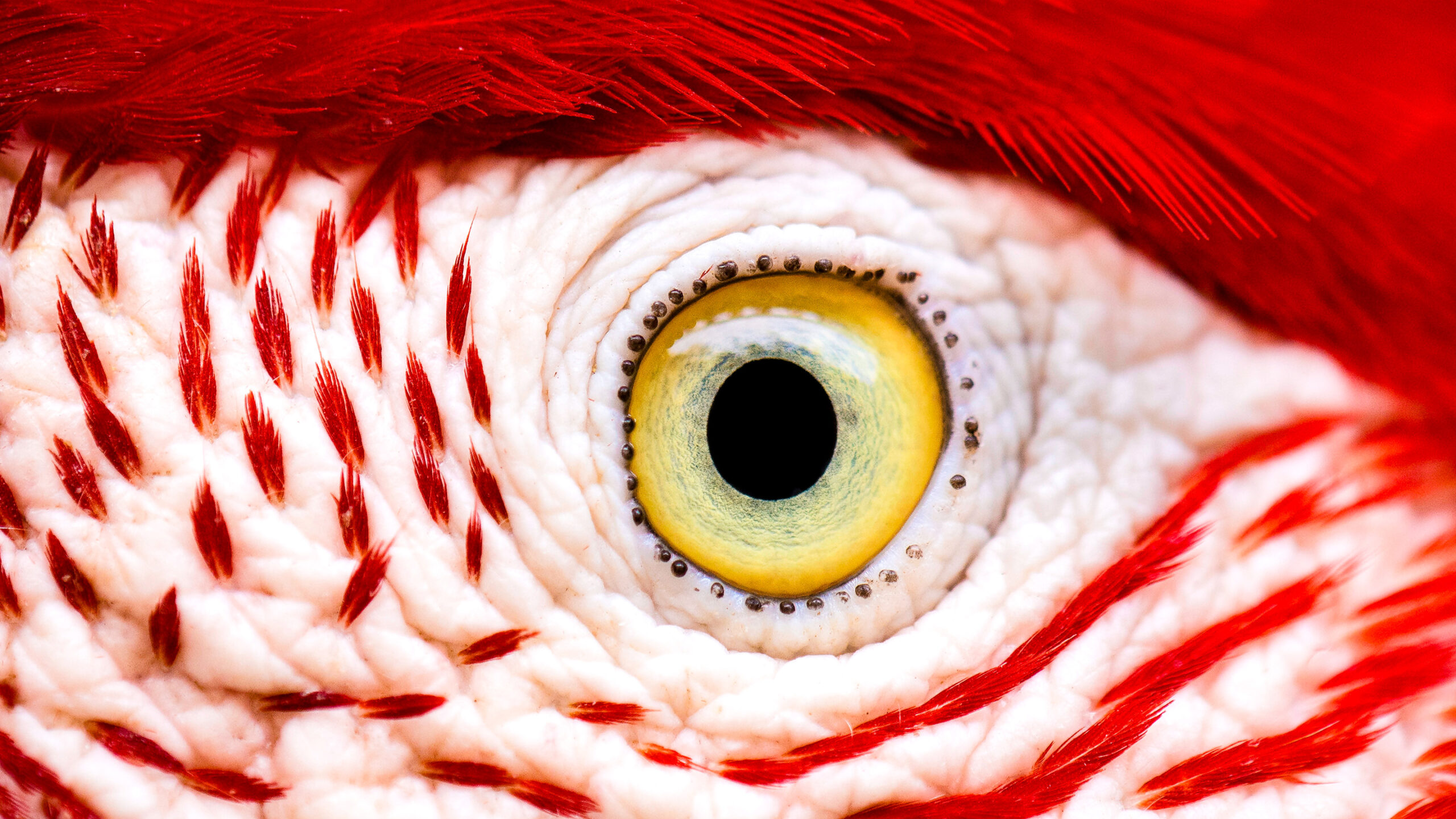
The eye of a red-and-green macaw, with no blood vessels in sight. How can a bird eye work so well without oxygen?
Leonardo Ramos
Introduction
When an optometrist shines a bright light into your eyes, a vast, branching tree sprouts in your field of vision. This is the shadow of blood vessels. Though we normally can’t perceive them, these vessels always occlude a portion of what we see, and for an important reason. They power the retina, a thin layer of nerve tissue in the back of the eye that communicates light signals to the brain.
The retina is one of the body’s most energetically expensive tissues. Built from complex networks of sometimes more than 100 different types of neurons, retinal tissue consumes two to three times more energy than the same mass of typical brain tissue. That’s why most vertebrate retinas, including our own, are furrowed with dense, branching networks of blood vessels: to deliver oxygen and other ingredients for producing energy.
But there’s a significant exception to this rule. Birds have retinas that mostly lack blood vessels. This may seem especially strange given birds’ exceptional vision. The bird retina is “one of the most metabolically active tissues in the animal kingdom, yet it worked with no apparent blood perfusion,” said Christian Damsgaard, an evolutionary physiologist at Aarhus University. “It was a complete paradox.” For centuries this has puzzled scientists, who figured that the bird retina must obtain oxygen through a unique, undiscovered process.
Damsgaard is the lead author of a study, published in the journal Nature in January 2026, that showed for the first time that bird retinas don’t have some unusual adaptation for acquiring oxygen — they survive without it entirely. Instead, to bring energy to the tissue, they use a process called anaerobic glycolysis that is significantly less efficient than oxygen-powered metabolism but gets the job done.

The evolutionary physiologist Christian Damsgaard measured gas exchange in bird eyes with microsensors. Surprisingly, the inner retina, a highly active tissue, used no oxygen.
Jesper Ekmann
By studying how tissues can survive without oxygen, researchers can potentially develop therapeutics to treat conditions of oxygen deprivation, such as strokes. More fundamentally, they want to understand the limits of evolution.
“What are the extremes of life?” Damsgaard said. “How far can we bend the conditions under which highly metabolically active tissues can actually survive?”
A bird, he learned, can bend them pretty far.
Oxygenated Life
Around 3.4 billion years ago, cyanobacteria invented photosynthesis. Slowly at first, then quickly, their newly evolved method of making energy from sunlight succeeded and spread. The cells pumped so much oxygen, a by-product of photosynthesis, into the atmosphere that it changed the course of life on Earth.
Oxygen molecules make energy production in cells extremely efficient. To extract energy, cells break down a glucose molecule into two pyruvate molecules. This process releases two molecules of ATP (adenosine triphosphate), life’s universal energy currency. A cell lacking oxygen can go only this far. Oxygen, however, enables further biochemical reactions that break down pyruvate and produce another 30 molecules of ATP. In other words, the presence of oxygen makes energy extraction from a single glucose molecule 15 times as efficient, and sometimes more.

Birds, such as this alpine chough (in the crow family), use their exceptional vision to hunt, forage, and migrate. This energetic ability is powered by an inefficient metabolism.
Jean-Paul Wettstein
The energetic advantage of oxygen, through the process of aerobic respiration, was transformative. Once oxygen imbued the atmosphere, evolution selected for organisms that could use it. “We’ve been hooked on 20% [atmospheric] oxygen for millions of years,” said Gary Lewin, a molecular physiologist at the Max Delbrück Center in Berlin. This Great Oxidation Event was followed by mass extinction, as organisms using oxygen outcompeted just about everybody else. While some life forms, such as certain bacteria, are adapted to life without oxygen, all complex, multicellular organisms need that energy advantage to survive.
Humans and most other animals can survive with little or no oxygen for several minutes at most. The mammal with the highest known tolerance for low-oxygen conditions is the naked mole rat, which can survive for up to 18 minutes breathing anoxic air in underground burrows. A few cold-blooded aquatic creatures, including freshwater turtles and goldfish, can persist in low-oxygen conditions at the bottom of a frozen lake for a year or two. But for most animals, a steady supply of oxygen is a must-have.
Without oxygen, a variety of processes shut down — especially in metabolically demanding tissues such as the brain. Without that energy, our cells malfunction and die.
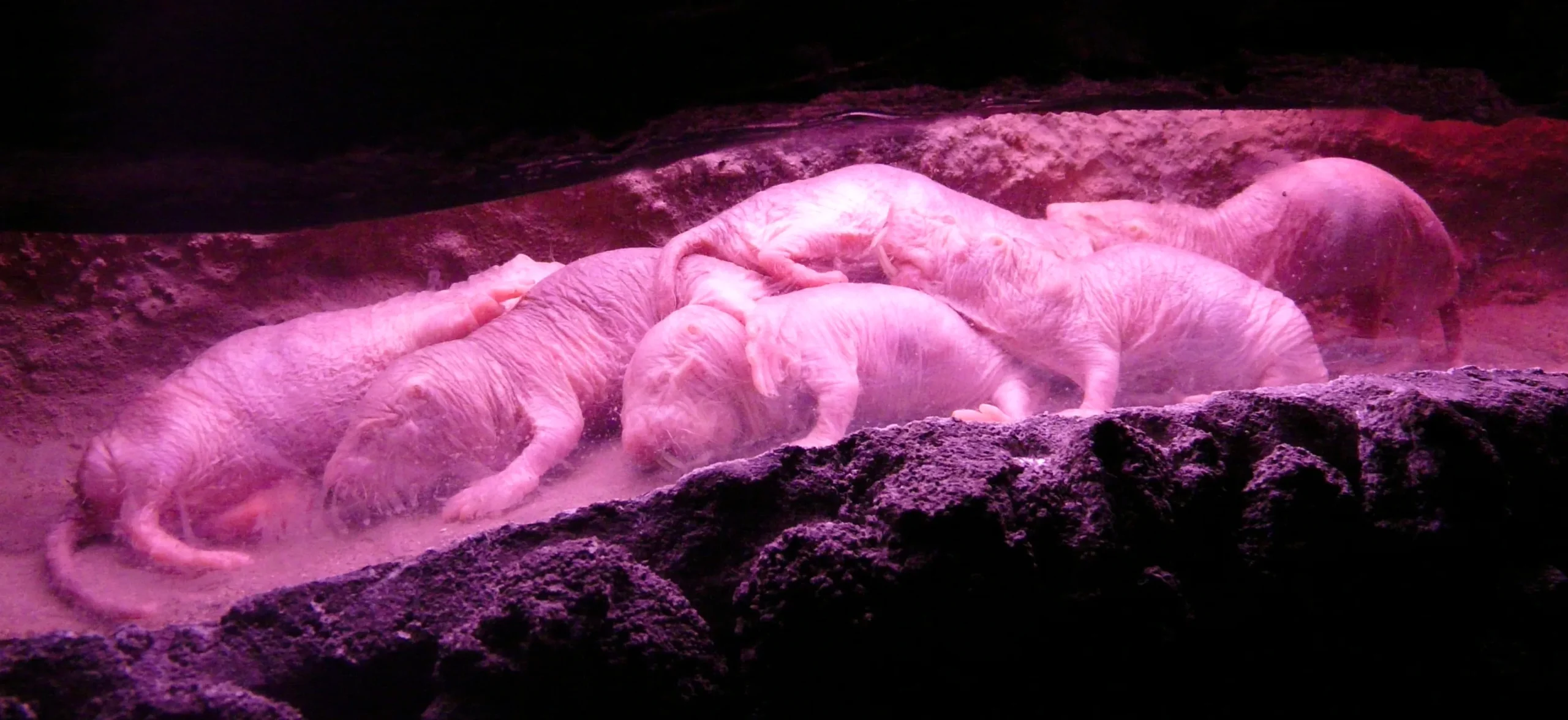
Naked mole rats can survive without oxygen for 18 minutes. To generate energy without oxygen, they use anaerobic glycolysis fueled by fructose.
Javier Ábalos
A Mysterious Structure
All this is why, in 2019, when Damsgaard learned that bird retinas lack blood vessels, he was confused. How could this high-energy tissue survive, let alone perform at the level observed in sharp-sighted bird species, without oxygen?
He pored over the voluminous research on the subject, all of which pointed at a mysterious structure in the bird eye known as the pecten oculi. In the 17th century, anatomists first described the unusual organ: It looked like a radiator, comblike, riveted with blood vessels, and with a large surface area. In the centuries that followed, researchers debated whether it helps deliver oxygen to retinal tissue in bird eyes. Damsgaard read about 30 different theories about the pecten oculi’s function based on anatomy alone.
“Nobody had really done direct physiological measurements on this structure,” he said. “That’s where we came in.”


Mark Belan/Quanta Magazine
In his lab, which studies the exchange of gases such as oxygen and carbon dioxide between vertebrates and their environments, Damsgaard’s team used microsensors to measure oxygen levels in the retinas of zebra finches, pigeons, and chickens. Indeed, in the inner retina, which completely lacks blood vessels, they found no oxygen. (They did measure oxygen in the outer retina, at the back of the eye, which has some blood vessels.)
That was “striking,” Damsgaard said. “Half of the retina lives in a chronic state of anoxia, where there’s no oxygen present at all.”
Using spatial transcriptomics, a method that combines cell imaging with RNA sequencing, the researchers mapped which genes were active in different parts of the retinal tissue. Genes associated with typical aerobic respiration were expressed in the outer retina, where there are blood vessels. In the oxygen-depleted inner retina, only genes associated with anaerobic respiration were active.
To trace the paths of nutrients, Damsgaard and his team worked with cancer scientists who are experts on oxygen-free metabolism (tumor cells often use anaerobic glycolysis to make energy). They found that the inner retina demanded 2.5 times more glucose than other parts of the bird brain.
Then they examined the pecten oculi. Their spatial transcriptomics data showed that the genes for glucose were highly active there. This suggested that the strange structure wasn’t bringing oxygen into the bird’s retina; rather, it was helping to pump glucose in, thereby enabling the less efficient anaerobic process.
As a by-product, anaerobic glycolysis creates lactic acid, which can accumulate and become toxic. The researchers also saw that genes for lactic acid transporters — the molecules that move lactic acid out of tissues — were active in the pecten oculi.
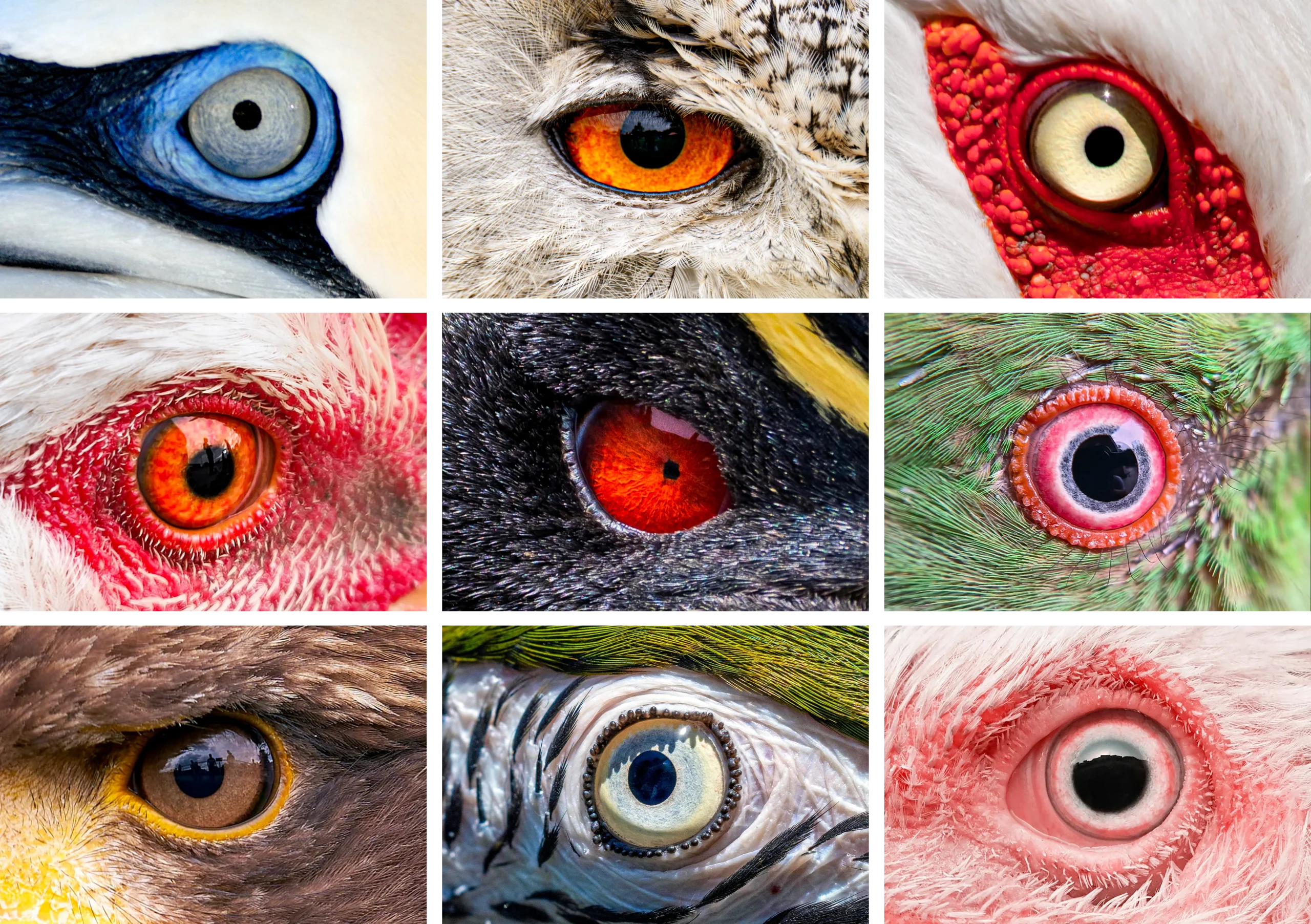
The diversity of bird eyes, lacking blood vessels (left to right). Top: Northern gannet, Eurasian eagle-owl, maguari stork. Center: rooster, rockhopper penguin, parrot (species unknown). Bottom: bald eagle, blue-and-yellow macaw, unknown species.
(Left to right) Top: Chris Hellier, Jiří Dočkal, Annette Lozinski. Center: Mohammed Brzan, Nico Marín, Shyamli Kashyap. Bottom: Ingo Doerrie, David Clode, Hasan Almasi
Their findings provide compelling evidence for the role of the pecten oculi in supporting anaerobic glycolysis, which “has been a mystery for a long time,” said Thomas Baden, a neuroscientist at the University of Sussex who was not involved in the study. “The insight that the retina basically goes oxygen-free, at least in some layers, is surprising. … It really gets properly down to zero.”
This pathway is used by cancer cells and temporarily by our muscles when they’re strained and can’t get enough oxygen — such as when we’re running. But no known vertebrate tissue was known to survive in completely anoxic conditions for a lifetime.
Eyes Like a Hawk
The bird’s retina and its no-oxygen power system are so unusual that they naturally raise questions about how they could have evolved.
This is “a series of beautiful experiments,” said Karthik Shekhar of the University of California, Berkeley, who was not involved in the research. It’s an example of how an animal took the vertebrate eye — a highly conserved structure whose origins go back some 560 million years to a light-sensitive patch on a primitive creature — and tinkered with it to fit its own needs. “Evolution is not really like an inventor; it acts more like a tinkerer,” he said, citing a 1977 essay, “Evolution and Tinkering,” by the French biologist François Jacob. “It takes parts that have existed long before, and it recombines, reinvents, and reshapes.”
The researchers tried to pinpoint when the pecten oculi might have arisen by comparing oxygen levels in the bird retina to those in not-so-distant relatives: two reptile species, Chinese pond turtles and broad-snouted caimans. The reptile retinas had normal oxygen levels and no indication of anaerobic glycolysis. This led Damsgaard’s team to conclude that the oxygen-free tissue likely evolved sometime during the dinosaur era, after the avian lineage had split from crocodiles but hadn’t yet evolved into modern birds. This was around the same time that the retina thickened.
Still, that rough time estimate can’t explain what evolutionary pressure might have selected for the unusual retinal tissue. Researchers can only speculate. “I think the system evolved in theropod dinosaurs in response to selection for sharp vision for tracking prey and identifying mates,” Damsgaard suggested. Then, later, when birds took to the skies, it “served as the physiological basis for maintaining retinal function” during high-altitude flights, when oxygen levels are low, he speculated.
The lack of blood vessels could also offer birds the advantage of better vision. The bird retina is complex and densely packed with more than a hundred cell types that work to render the world in great resolution. Birds use their exceptional visual sense for hunting and foraging — consider an owl tracking a mouse from the sky, an albatross watching for signs of fish on the ocean’s surface, or a hummingbird locating hundreds of flowers every day — as well as for following landmarks across the landscape during migration. Without blood vessels obstructing their view, birds’ retinal cells might be able to take in more visual information.
Could this be an adaptation, or is it a coincidence of evolutionary history? There’s no way to know for sure how birds’ incredible vision evolved. There’s this mystery “that has lingered around us,” Baden said. “What is it about birds that makes their eyes so special?” Their retinal power system seems as if it could explain what makes them so unique. However, Lewin, the physiologist, is cautious about overextending the results and interpretations to every bird, given that the researchers haven’t looked at any migratory species.
The implications stretch well beyond bird adaptations to biomedicine. A common thread in many medical conditions is a drop in oxygen delivery to tissues, which, depending on where it occurs, can lead to scars or brain damage. Human brains can tolerate maybe a minute of total anoxia, Lewin said. That’s what makes strokes, which cut off blood and oxygen supply to parts of the brain, so devastating. By studying low-oxygen conditions in creatures such as naked mole rats and birds, scientists can gain insight into how tissues can tolerate low-oxygen conditions.
“Maybe we can get inspiration for how nature solved these problems by millions of years of natural selection,” Damsgaard said. “There’s so much to be learned from these animals that are able to do something that we cannot do.”